A new study has helped researchers from several universities understand how nerve cells behave when recording memories of a place or of a person. A brain area known as the medial temporal lobe turned out to be essential for the process of memory formation.
One of the many benefits of the study, published earlier this week, on Wednesday (July 1, 2015), in the journal Neuron, is that it might help experts learn how memory loss and memory formation differ in patients with neurological disorders such as Alzheimer’s.
The scientific community has been trying to solve the mystery of how the brain forms memories for many years. Some of the previous studies have theorized that wither solitary nerve cells, or just handful of them are responsible for representing a person or a place. Other theories go in the opposite direction, saying that memories require a great number of neurons in order to be formed.
One of the most promising studies conducted in the past, and the one that inspired this new study, found that there is a neuron, dubbed “the Jennifer Aniston neuron”, which activated every time a person looked at images of the celebrity.
It is worth mentioning that Prof Rodrigo Quian Quiroga was involved in the “the Jennifer Aniston neuron” study, and used it as inspiration for the new study, which he collaborated on with Dr. Matias Ison from the University of Leicester and with various scientists from the University of California.
The project involved selecting 14 people who suffered from severe epilepsy and hooking them up with electrode implants so that the researchers can monitor which areas of the brain host the seizures. The implants also served to help experts detect individual neurons responsible for forming and storing memories.
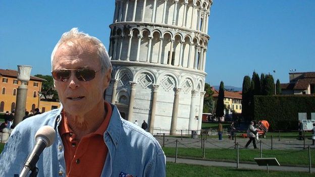
The subjects were presented with roughly 100 photos of people and places. Among the human faces, celebrities such as Clint Eastwood, Julia Roberts, Jennifer Aniston, Halle Berry and Tiger Woods could all be recognized. Images of famous places included the Eiffel Tower, the White House and Leaning Tower of Pisa.
The researchers first had subjects look at the photos of celebrities, and noticed that there were specific nerve cells which activated when they saw each of the faces. The team than had the subjects look at the photos of places, all of them unrelated to the celebrities. They notice that there were other specific nerve cells which activated when people saw this second set of images.
The final step of the test was to digitally merge together the two (2) separate sets of images. For instance, Clint Eastwood was photoshopped in front of the Leaning Tower of Pisa.
After showing the subjects the digitally modified images, the researchers went back to showing them the images that portrayed just a place, without a person. The experts reported observing that nerve activity changed drastically here as the subjects had already learned that there was a new connection between a person and a place.
What happened was that while subjects were looking at images of just the Leaning Tower of Pisa, the neuron which previously activated when seeing the Tower, as well as the one which previously activated when seeing Clint Eastwood, both responded to the image.
Dr. Itzhak Fried, neurosurgeon over at the UCLA Medical Center as well as the University of California’s Geffen School of Medicine, gave a statement explaining that “When the association is created, suddenly the cell very rapidly changes its firing properties”.
Dr. Matias Ison also gave a statement, sharing that this is the first study to explore how a single, solitary neuron correlates learning new contextual links that form in the brain.
Image Source: bbci.co.uk
Leave a Reply
You must be logged in to post a comment.