Example of tunable patterning
The invention of 3D printing has generated a huge advance in technology, allowing for a plethora of brand new devices being invented. The technique can even be used in medicine, with people actually getting 3D printed limbs. But cellular precision is very difficult to get, so researches from three different universities developed a way to move individual cells around in 3D with acoustic tweezers.
- The project is a collaboration between three different universities
- The paper was published in the journal Proceedings of the National Academy of Sciences
- A human heart contains over 2 billion muscle cells
- Acoustic tweezers can harmlessly move around, remove, and even transport any single cell
- Pretty much all fields of medicine would benefit from the technology
A team formed of researchers from Carnegie Melon, MIT, and the Pennsylvania State University is responsible for devising the highly sensitive and accurate technology.
The technique called acoustic tweezers uses very precise sound waves to trap and manipulate each individual cell, without any need for invasive contact.
It causes no cellular damage whatsoever, and the team used to move and play around with cells in a wide variety of waves in order to prove its efficiency and harmlessness.
The main purpose behind the invention is to help in the 3D printing of biological material – bioprinting, if you will.
Because of the insane amount of microscopic cells present in any one organ or body part, scientists need a way to extremely precisely move cells around so that no mistake is made in the finished product.
If any single cell is misplaced, is can lead to whole array of different health complications, including cancer.
By developing the acoustic tweezers, the tri-university team of researchers ensures the proper development of bioprinting technology.
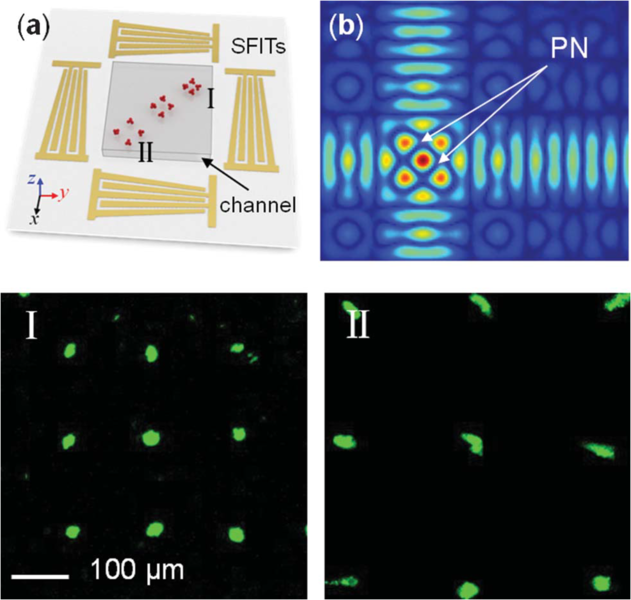
The most recent incarnation of the acoustic tweezers device involves a microfluidic part that employs acoustic wave generators in order to create sound waves along the device’s edges.
Due to the device’s design, the team was able to manipulate how sound waves moved along all three axes, at their meeting point forming a sort of cell-capturing node. From there, they could do with the cells whatever they wanted, without fear of harming them.
To further demonstrate the device’s capabilities, the team used the device to place the collected cells in a previously determined pattern, showing an incredible control over the cells’ placement, geometry, and spacing.
The next step is to employ the device in actually bioprinting a functional organ, while future applications would help the fields of bio manufacturing, cancer, neuroscience, tissue engineering, and even regenerative medicine.
Image source: Wikimedia
Leave a Reply
You must be logged in to post a comment.